

Early kidney dysfunction often develops without obvious symptoms. Patients may feel well while microscopic changes begin within the glomeruli and renal tubules. By the time serum creatinine rises or estimated glomerular filtration rate declines, structural injury may already be present.
A urine test for kidney function provides a direct window because the kidneys continuously filter blood and produce urine. Subtle shifts in protein excretion, albumin leakage, or urinary sediment can appear long before blood markers change. When these measurements are performed using FDA-cleared quantitative urinalysis rather than semi-quantitative dipstick methods, clinicians gain more precise data for early detection.
Why Early Detection Matters
Chronic kidney disease progresses in stages. In early phases, damage may be limited to mild glomerular permeability changes or tubular dysfunction. Intervention at this point can slow progression and reduce long-term complications.
Common risk factors for early renal dysfunction include:
- Diabetes mellitus
- Hypertension
- Cardiovascular disease
- Autoimmune disorders
- Long-term use of nephrotoxic medications
In these populations, small urinary biomarker changes may represent the first measurable sign of renal involvement. Detecting these changes requires sensitive and reproducible testing methods.
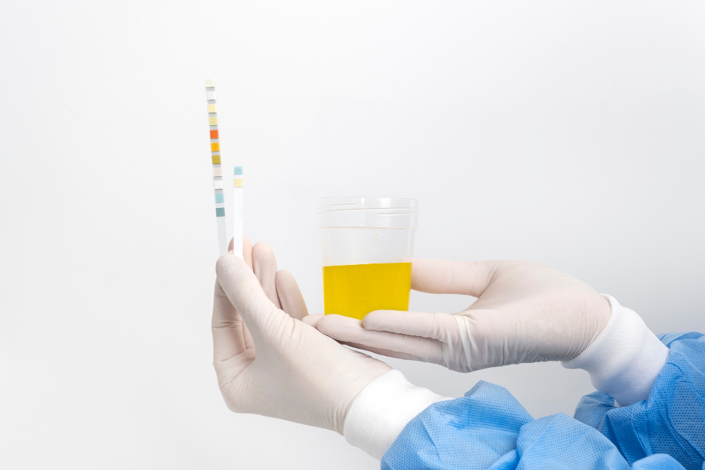
Limitations of Traditional Dipstick Screening
Dipstick urinalysis has long been used for rapid screening. While convenient and inexpensive, dipsticks provide semi-quantitative categories such as negative, trace, 1+, or 2+. These categories are based on colorimetric reactions that depend on urine concentration and visual interpretation.
Several limitations affect early detection:
- Detection thresholds may miss low-level albumin excretion
- Visual interpretation introduces operator variability
- Concentrated or dilute urine alters color intensity
- Interfering substances can affect reactions
Because early kidney dysfunction often presents with subtle albumin or protein increases, dipstick screening may not detect meaningful abnormalities.
Quantitative urine testing addresses these limitations by providing numeric measurements rather than broad categories.
Albumin: An Early Marker of Glomerular Injury
Albumin is one of the most sensitive indicators of early glomerular damage. Under normal conditions, only small amounts of albumin pass into urine. Increased permeability allows greater albumin leakage, even before overt kidney failure develops.
Quantitative urine testing measures albumin concentration precisely. This allows detection of low-level elevations that fall below dipstick detection thresholds.
In clinical practice, albumin measurement is often paired with creatinine normalization. The albumin-to-creatinine ratio accounts for urine dilution and provides a standardized value for interpretation.
Small increases in this ratio may indicate early nephropathy, particularly in diabetic or hypertensive patients. Identifying these changes allows earlier therapeutic intervention.
Total Protein and Tubular Dysfunction
While albumin reflects glomerular permeability, total protein measurement captures additional proteins that may signal tubular injury or inflammatory processes.
Quantitative protein analysis provides:
- Objective numeric concentration
- Improved sensitivity compared to dipstick testing
- Reliable trending over time
Mild but persistent protein elevation may indicate early renal stress. When tracked longitudinally, gradual increases may signal progression before other laboratory abnormalities appear.
Automated urine chemistry systems provide reproducible measurement across operators and shifts, reducing variability that could obscure subtle trends.
Creatinine Normalization and Dilution Effects
Urine concentration varies based on hydration, diet, and time of day. A concentrated specimen may show higher analyte levels simply due to reduced water volume. Conversely, dilution may mask abnormal excretion.
Creatinine normalization addresses this issue by expressing analyte concentration relative to urinary creatinine. This approach reduces dilution-related distortion.
For example:
- Albumin concentration divided by creatinine concentration
- Ratio expressed as mg albumin per g creatinine
This normalized value allows more accurate comparison across visits and patient populations.
In early kidney dysfunction, slight increases in albumin-to-creatinine ratio may represent the first measurable abnormality.
Microscopic Changes and Particle Analysis
Quantitative urinalysis is not limited to chemistry markers. Automated particle analysis provides standardized counts of urinary sediment elements, including:
- Red blood cells
- White blood cells
- Epithelial cells
- Casts
- Crystals
Microscopic hematuria may indicate glomerular inflammation or structural abnormalities. Increased white blood cells can reflect inflammatory or infectious processes affecting the urinary tract.
Automated particle quantification improves reproducibility compared to manual microscopy. Digital imaging systems apply standardized classification criteria, reducing subjective variability.
Integrating chemistry and particle findings strengthens early detection of renal abnormalities.
Trending for Long-Term Monitoring
Early kidney dysfunction rarely presents as an abrupt change. Instead, biomarker levels may shift gradually over months or years.
Quantitative urine testing allows clinicians to:
- Identify incremental increases in albumin excretion
- Monitor response to antihypertensive or antidiabetic therapy
- Compare results across visits
- Detect subtle progression
Numeric reporting provides continuous data rather than categorical results. This enables more refined interpretation and promotes proactive management.
Trend analysis is particularly valuable in chronic disease monitoring, where small shifts may have long-term significance.
Reducing False Positives and False Negatives
Semi-quantitative dipstick methods are influenced by urine pH, concentration, and chemical interference. These factors can produce inaccurate readings.
Quantitative urine chemistry systems measure analytes using validated methodologies that reduce subjectivity. Standardized calibration and automated analysis improve reproducibility.
By minimizing variability, quantitative testing increases clinical confidence in reported values. This reliability is particularly important when evaluating small deviations from baseline.
Workflow and Reporting Advantages
Modern laboratory systems integrate quantitative urinalysis into electronic workflows. Automated platforms measure multiple analytes in a single run and transmit results directly through laboratory information systems.
Advantages include:
- Objective numeric output
- Automated ratio calculation
- Standardized reference intervals
- Reduced manual handling
This integration benefits both high-volume laboratories and point-of-care environments. Consistent reporting improves clarity for clinicians reviewing results.
Improved workflow efficiency also allows laboratory staff to focus on cases requiring additional review or correlation.
Identifying High-Risk Patients Earlier
The greatest benefit of quantitative urine testing lies in its ability to detect renal abnormalities at an earlier stage.
High-risk populations benefit from routine monitoring, including:
- Patients with diabetes undergoing annual screening
- Individuals with long-standing hypertension
- Patients receiving medications known to affect renal function
Small but measurable changes in urinary biomarkers may prompt medication adjustments, lifestyle counseling, or referral to nephrology services.
Without quantitative measurement, these early warning signs may remain undetected.
Detect Renal Changes Earlier with AutoUA
Early kidney dysfunction requires precise, reproducible measurement. AutoUA is the only FDA-cleared quantitative urinalysis system that replaces traditional dipstick testing with objective numeric results. Designed for compatibility with leading clinical chemistry analyzers, AutoUA integrates urine chemistry, particle analysis, and creatinine measurement into a fully automated workflow.
With LIS connectivity and standardized reporting, laboratories can strengthen diagnostic confidence while improving efficiency.
Speak with our technical team to learn more, request pricing information, or schedule a live demonstration to see how AutoUA can support earlier renal detection in your facility.